Local elementary school (5th graders) – plant
cells and
animal cells with Dr.
Chris Sacchi (and me, of course!)
The 1st and 5th
graders from the local elementary school visited
Kutztown University to learn the difference betwee plant and animal
cells using the Microscopic Digital Imaging Lab.
The questions of the
day were:
1) Are plants and animals really made of cells?
2) How do animal cells and plant cells differ?
The microscope we used had a
digital camera attached to it so we could take photographs of the
different cells for comparison.
We first looked at plant
cells.
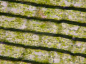 The
image to the
right shows plant cells from the elodia
plant - a floating, flowering
plant
common in ponds that has leaves that are only two cells thick!
The green specks are chloroplasts, which are the little packets inside
the cells containing chlorophyll (the molecule that allows plants to
convert sunlight + carbon dioxide + water into sugar).
The
chloroplasts float around in the cell fluid (called cytoplasm) and try
to orient themselves so that they are exposed to as much light as
possible. Click
here to see a 6 Mb Quicktime® movie of these elodia plant
cells experiencing "cytoplasmic
streaming." The
cell is sort of like a vegetable stew, slowly convecting and churning.
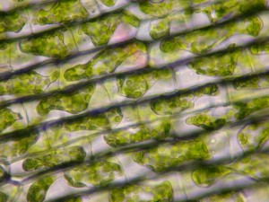 Most of the cell parts (called
organelles) are almost invisible because they are colorless. Dr.
Sacchi put a little salt water on the slide to see what would
happen. The insides of the cells squished up and the chloroplasts
all bunched together because the water in the cytoplasm was "sucked
out"
of the cell by a phenomenon called "osmosis."
Most of the cell parts (called
organelles) are almost invisible because they are colorless. Dr.
Sacchi put a little salt water on the slide to see what would
happen. The insides of the cells squished up and the chloroplasts
all bunched together because the water in the cytoplasm was "sucked
out"
of the cell by a phenomenon called "osmosis."
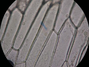 We
also looked at the cells of an onion bulb. Since the onion bulb
grows underground, it doesn't see any sunlight and so it doesn't have
any chloroplasts for photosynthesis. Not having all those
chloroplasts in the way, we wanted to see the nucleus of the cell,
which is where the DNA is stored. We added a drop of iodine to
the slide, which acted as a stain that made the nucleus visible.
The little circular dot inside each cell is the nucleus.
We
also looked at the cells of an onion bulb. Since the onion bulb
grows underground, it doesn't see any sunlight and so it doesn't have
any chloroplasts for photosynthesis. Not having all those
chloroplasts in the way, we wanted to see the nucleus of the cell,
which is where the DNA is stored. We added a drop of iodine to
the slide, which acted as a stain that made the nucleus visible.
The little circular dot inside each cell is the nucleus.
 Now it was time to compare plant
cells with animal cells, but where
would we find an animal?
Now it was time to compare plant
cells with animal cells, but where
would we find an animal?
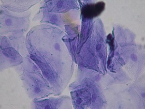
We found an animal by looking in the mirror!
Using
a toothpick, we scaped some cells off the inside of our cheeks.
You can imagine how often the skin on the inside of your cheeks rubs
against your teeth -- whenever you move your mouth! Every time
you move your mouth, a few cheek cells (called epithelial cells) rub
off and you swallow them.
That made me think. The cells are very small, so we don't swallow
very much each time, but if I were to add up all of the times that I
have swallowed in my whole life, I wonder if all of those the swallowed
cheek cells would add up to be as big as my whole body!
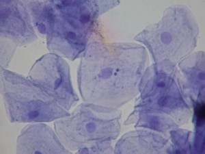 We
stained the cells using a chemical called methylene blue, which reacts
with acids to turn purple. Those parts of the cell that contained
some acid turned purple. The soft purple circles in each cell are
the cell nucleus - why do you suppose the nucleus stained purple?
What molecules are acids that you would find there? (Hint: the
molecules are shaped like a double helix!)
We
stained the cells using a chemical called methylene blue, which reacts
with acids to turn purple. Those parts of the cell that contained
some acid turned purple. The soft purple circles in each cell are
the cell nucleus - why do you suppose the nucleus stained purple?
What molecules are acids that you would find there? (Hint: the
molecules are shaped like a double helix!)
You can also see little dark purple dots on the surface of the
cells. Those are bacteria! Everyone has billions of
bacteria growing in their mouths and all over their bodies! It's
natural. Some of those bacteria are actually good for us - they
help us in many ways and all they ask is for a little place to live!
So, do you see any differences between plant and animal cells?
I'm a teacher, so you didn't think I was going to just tell you the
answer right away, did you? My job is to set people up learn
things for themselves!
You have the pictures - study them and you can figure it out!
Everybody had a chance to use the
microscopes - it was a fun day for all!
 The
image to the
right shows plant cells from the elodia
plant - a floating, flowering
plant
common in ponds that has leaves that are only two cells thick!
The green specks are chloroplasts, which are the little packets inside
the cells containing chlorophyll (the molecule that allows plants to
convert sunlight + carbon dioxide + water into sugar).
The
image to the
right shows plant cells from the elodia
plant - a floating, flowering
plant
common in ponds that has leaves that are only two cells thick!
The green specks are chloroplasts, which are the little packets inside
the cells containing chlorophyll (the molecule that allows plants to
convert sunlight + carbon dioxide + water into sugar). 


